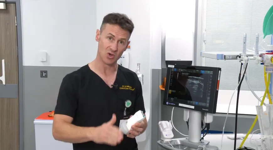
ICU and Anaesthesia Consultant Dr. Jonny Wilkinson demonstrates how to carry out both pelvic view and abdominal aorta views using ultrasound scanning.
Firstly Dr. Wilkinson shows the pelvic view on abdominal scanning, looking at the bladder by rotating the probe so that the marker is at the patient's left-hand side and the beam angled down over the brim of the pelvis.
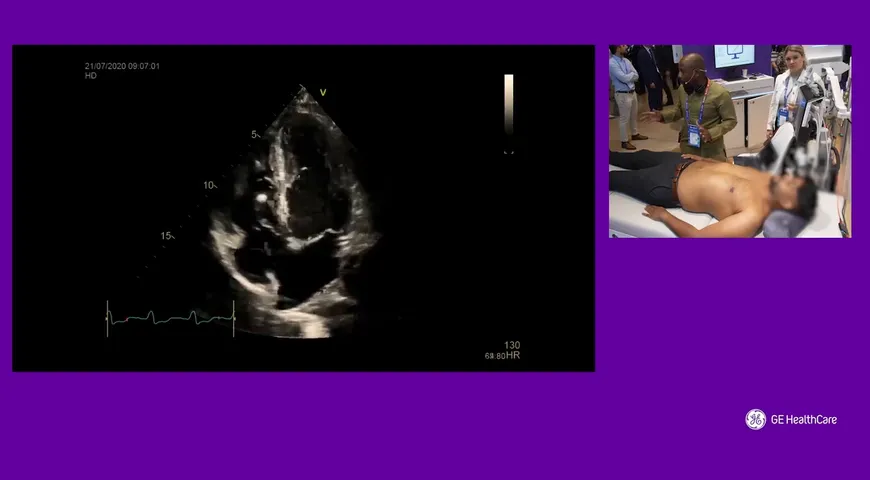
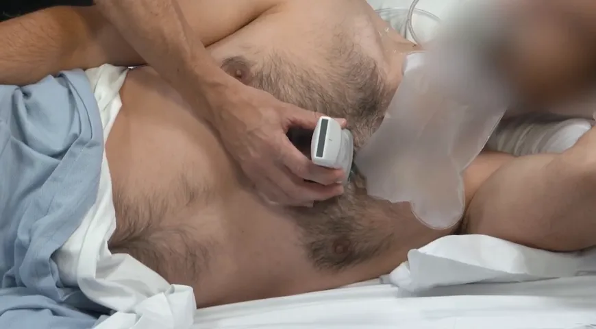
He then goes on to demonstrate the view of the abdominal aorta. Positioning the curvilinear probe approximately two centimetres just above the patient's umbilicus, with the marker at the patient's left-hand side, aiming to see the vertebral body shadow at the back and the pulsation of the abdominal aorta on the screen.