Dr. Belinda Chan, M.D. is an Assistant Professor in the Division of Neonatology, Department of Pediatrics at the University of Utah, School of Medicine. Dr. Chan was an early evaluator participant of the product. In this video you will listen to the daily challenges faced and how POCUS is used within the NICU to improve patient outcomes. A concern with NICU patients is lung, using lung ultrasound helps to provide a differential diagnosis. The Venue Family lung Diagram< saves Dr. Chan time due to the auto labelling plus acts as a reminder to capture all images to give an overview of the patients lung. Listen to the numerous uses of POCUS in the NICU, from umbilical venous and artery catheter placement, bladder assessment, lumbar puncture and spinal tap procedures.
Join us and listen to how the Venue Family can benefit your POCUS when working within the NICU.
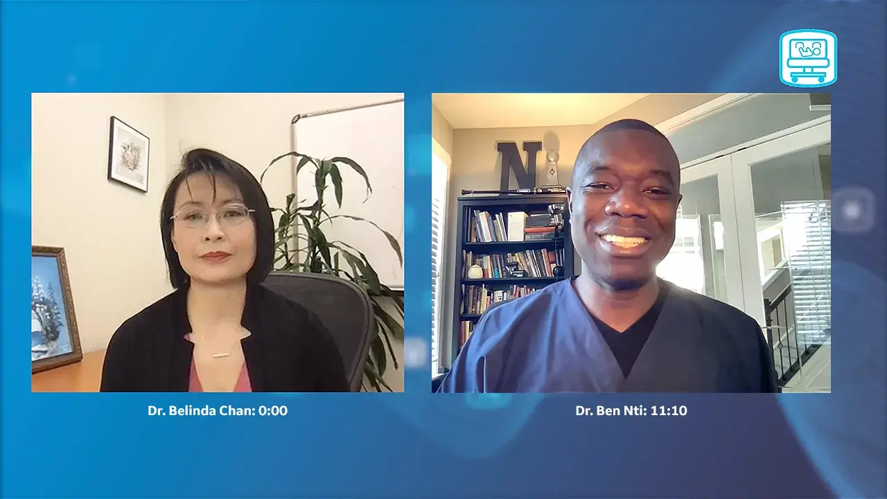
Dr. Ben Nti, MD is an assistant Professor of Emergency Medicine, Indiana University School of Medicine Indianapolis, U.S.A.. Dr. Nti developed and interest in Pediatric Point of Care Ultrasound (POCUS). Dr. Nti was an early evaluator participant of the product. Dr. Nti gives an overview of the most common pediatric conditions he faces on a daily basis. One particular area of discussion is Lung ultrasound. Dr. Nti discusses how the newly released clinical tools on the Venue Family help him address these pediatric conditions, specifically lung. The first tool discussed is the Lung Diagram and how this allows him to ensure he covers each segment of the lung giving a whole picture of the lungs rather than searching for the individual images. The second tool discussed is Lung Sweep, listen to how Dr. Nti feels this tool helps him to diagnose pneumonia at the patient beside, as the panoramic view allows him to view all the pleural in 3 views, Anteriorly, Axillary and Posteriorly. The Venue Family also has 2 further diagrams which have been beneficial to Dr. Nti and his team, the renal diagram and the eFAST diagram.
Join us and enjoy listening to how the Venue Family can be of benefit to your daily POCUS when dealing with pediatrics.
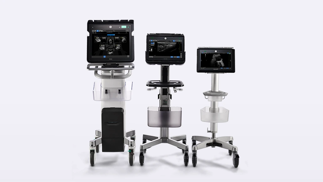
Learn more