Made for your point of care

Made for critical moments

Dr. Jonathan Wilkinson
Consultant in Intensive Care (UK)

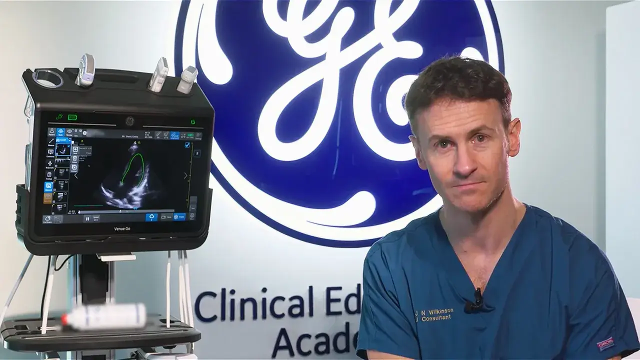
Dr. Wilkinson is an Intensive Care and Anesthesia Specialist Consultant at Northampton General Hospital where he now is involved in leading the intensive Care Unit, providing Consultant led safe anesthesia to elective and trauma patients and attending cardiac arrests, trauma calls and ward based peri-arrest situations. Dr. Wilkinson is an avid user of point of care ultrasound and is heavily involved in point of care ultrasound trainings as a focused intensive care echocardiography mentor. Dr. Wilkinson was an early evaluator participant of the product.

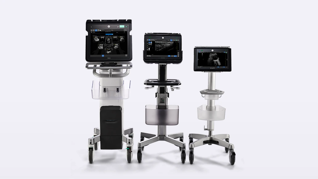
This video shows Dr. Wilkinson discussing the newly released clinical tools on the Venue Family (Venue and Venue Go) and why these Point of Care ultrasound tools help him during his assessment of the ICU patient. He will also demonstrate Lung ultrasound during live-scanning sessions, using the new L4-20t-RS XDClear™ probe.

The first new tool discussed is Lung Sweep including the existing Lung Diagram and B-Lines tool. Dr Wilkinson believes the combination of these tools is going to revolutionize the way he looks to his patients with ultrasound, more particular from the lung ultrasound angle.

The Lung Sweep tool creates a dynamic panoramic view of the lung (Anterior, Lateral, Posterior per left/right side). These views allow 2D coronal and sagittal coverage of the entire lung parenchyma which assists him in the diagnosis of pneumonia.

Real Time Ejection Fraction (Real Time EF) is another new tool to the Venue Family (Venue and Venue Go). Real-Time EF is an AI enabled tool that continuously calculates Real-Time ejection fraction during live scanning in apical 4CH view and allows users to capture instant, precise results. Dr. Wilkinson will demonstrate how the tool works on a live model.

Join us and enjoy listening!

Learn more
The Venue™ Family

Made for your point of care.

Device Images

3 systems. 1 Shared platform.

Designed for your needs, adapts for your challenges.

AI Images GIF

Precise AI tools, designed to enable fast patient assessments and confident decisions.

Work smarter, not harder.

Related videos

Made for Anesthesia

Made for anesthesia

Dr. Amit Pawa

Made for NICU and Pediatrics

Made for NICU and pediatrics

Dr. Belinda Chan & Dr. Ben Nti

Made for MSK

Made for MSK

Dr. Fernando Jiménez Díaz

White papers

Auto Tool for Measuring IVC Collapsibility Index

Auto Tool for Measuring IVC Collapsibility Index

Auto Tool for Measuring VTI

Auto Tool for Measuring VTI

Lung Ultrasound for Assisting in the Diagnosis of Pneumonia

Lung Ultrasound for Assisting in the Diagnosis of Pneumonia

Real-Time Documentation

Real-Time Documentation

Real-Time EF

Real-Time EF

SonoDefense on the Venue™ Family

SonoDefense on the Venue™ Family

Questions & Answers

How long does it take to obtain the EF with the new AI-tool?

The Venue Family offers a novel semi-automated Real-Time EF tool. The EF may be obtained within 3 seconds of live scanning in A4CH view, when image quality is sufficient as determined by the Quality Indicator.

What is Lung Sweep?

The Velocity Time Integral (VTI) measurement plays a significant role in the management of critically ill patients. It allows the physician to monitor hemodynamic changes resulting from a disease or from the therapy actions that were required to achieve better patient outcome.1

What is the frequency of the L4-20t-RS?

L4-20t-RS XDclear™, is an ultra-wide band probe from 4-20MHz supports high-frequency imaging of superficial structures while also being able to penetrate deeper anatomy without compromising imaging quality.

Is ECG needed for the Real Time EF tool to work?

No ECG is required for the tool to work.

What is the difference with the current EF automated tools?

The Venue Family Real-Time EF is an AI enabled tool that continuously calculates Real-Time ejection fraction during live scanning in apical 4CH view and allows users to capture instant, precise results.

What are the Easy & fast exam documentation?

Easy documentation features provide a visual overview of the entire exam while reducing the need to manually type findings. Exams are stored as one single image and users can easily switch between exams with one simple click. We have Lung Diagram / eFAST and Renal Diagram.

How VTI is used for patient management?

  1. VTI trending can be used to assess response to therapy.
  2. Assessing the absolute values of VTI may be useful to estimate cardiac output, assessing the relative change of VTI in shock management may be practically more useful to assess the response to therapy and help in decision making process about the next therapeutic steps.
  3. If SV / VTI increases by 15% with fluid challenge fluid responsive may be considered.2
  4. If SV / VTI increases by 20% with inotropes contractile reserve may be considered.3
1. Auto VTI can provide up to 90% reduction in keystrokes and take up to 82% less time than manual method calculations, as performed by experts. Based on GE Internal study with Venue Go DOC2254811.
2. A recent study found the Auto B-lines tool to be comparable to and as highly reliable as visual counting performed by experts. ShortJ, Acebes C, Rodriguez-de-Lema G, et al. Visual versus automatic ultrasound scoring of lung B-lines: reliability and consistency between systems. Med Ultrasonography 2019, Vol 21 no1, 45049 DOI: 10.11152/mu-1885.
3. Supporting evidence for Venue and Venue Go is documented in DOC2391130. Supporting evidence for Venue Fit is documented in DOC2454794.