 Clinical specialty
 Clinical specialty
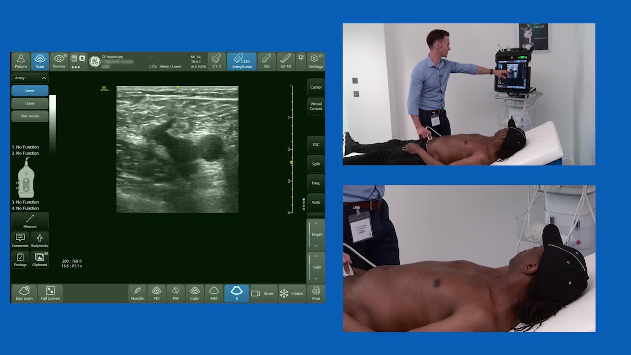
Watch a video demonstration of Dr. Jonny Wilkinson (MBChB MRCP FRCA FFICM) performing ultrasound scanning to assess the saphenofemoral junction and determine deep vein thrombosis.
When a blood clot forms in an artery or a vein and restricts your blood flow, it is known as thrombus; deep vein thrombosis (DVT) is a blood clot that is formed deep in one of your veins in your body, most commonly in one of your legs.
Ultrasound scanning is a highly accurate tool to use to diagnose DVT as Dr. Wilkinson explains in the video. Firstly he places the ultrasound probe on the saphenofemoral junction, which is located near your groin. The ultrasound operator will be able to compress your great saphenous vein to identify a blood clot.
In this video, Dr. Wilkinson shows the great saphenous vein compressing and therefore eliminating DVT from the patient. Click the button to watch now.

Dr. Thibaut Markarian

Prof. Laurent Muller

Prof. Laurent Muller

Dr. Thibaut Markarian

Prof. Laurent Muller

Prof. Laurent Muller
| Provider | Cookies |
|---|---|
| POCUShub | cookieconsent,token,healthcarepro,tab,PHPSESSID |
| Marketo | __cf_bm |
| Provider | Cookies |
|---|---|
| NID,CONSENT,1P_JAR,CONSENT,ANID,NID | |
| Doublick.net | IDE |
