Social wall

How to assess venous congestion

Assessment of venous congestion

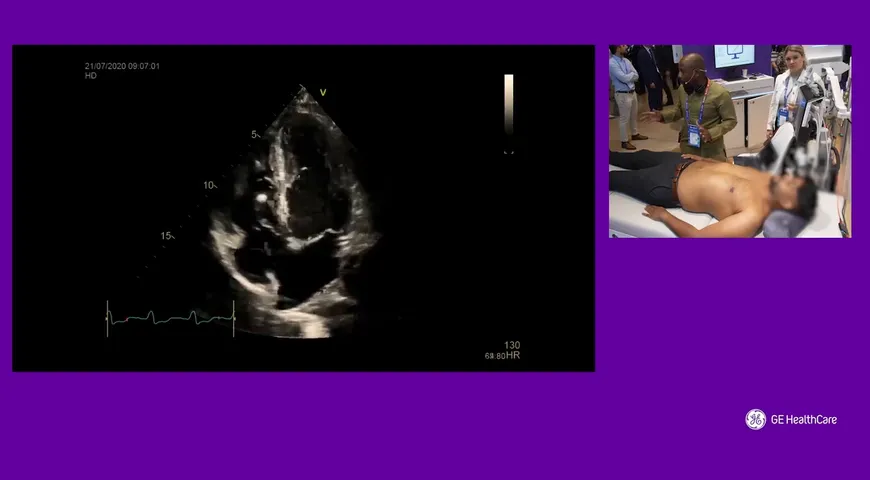
Dr Segun Olusanya is an intensive care specialist trainee from London talk us through the most effective process of assessing venous congestion on a patient.
Venous congestion refers to a common complication of traumatic injuries and reconstructive surgery. Venous congestion occurs when arterial inflow is greater than venous outflow. When venous outflow is obstructed by clotting or disruption of veins, venous pressure increases.

The starting point for this process is the inferior vena cava using a cardiac probe, he then discusses the best practice to perform an assessment from a venous congestion perspective. If the inferior vena cava is more than 2.1cm your patient may have venous congestion and you will need to start looking at other signs, Segun helps us become aware of what signs are relevant to this diagnosis.
To learn more about Venous congestion, continue watching the video below.

Related

Empowering Critical Care with Ultrasound: A Hands-On Demonstration by Dr Olusegun Olusanya
37:22 min

Empowering Critical Care with Ultrasound: A Hands-On Demonstration by Dr Olusegun Olusanya

Empowering Clinical Practice: Jonny Wilkinson Explores the Tools of the Venue Sprint
15:33 min

Empowering Clinical Practice: Jonny Wilkinson Explores the Tools of the Venue Sprint

Dr. Jonny Wilkinson

Optimising Critical Care with Ultrasound: Jonny Wilkinson on the Venue Sprint
06:20 min

Optimising Critical Care with Ultrasound: Jonny Wilkinson on the Venue Sprint

Dr. Jonny Wilkinson

Lung Ultrasound Pearls
01:28 min

Lung Ultrasound Pearls

Dr. Ashley Miller

Contrast-Enhanced Ultrasound- Guided Biopsies at Odense University Hospital
04:03 min

Contrast-Enhanced Ultrasound- Guided Biopsies at Odense University Hospital

Prof. Christian B. Laursen

L’évaluation des fonctions VD et VG : aller plus loin que l’évaluation visuelle.
41:39 min

L’évaluation des fonctions VD et VG : aller plus loin que l’évaluation visuelle.

Prof. Laurent Muller