How to assess the right ventricle

Assessment of the right ventricle

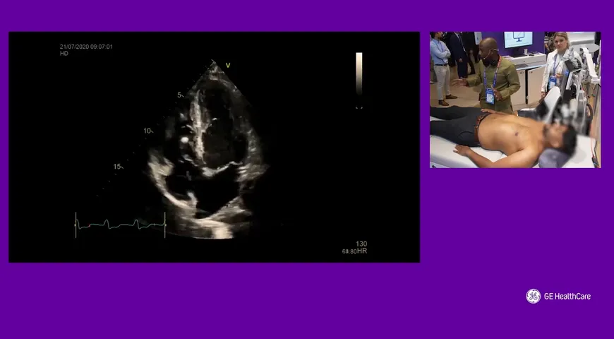
Dr Marcus Peck, a consultant intensivist based in Firmley Park Surrey.  He gives us an insight into how to assess the right ventral of the heart, which is the lower right chamber of the heart that receives deoxygenated blood from the right atrium and pumps it under low pressure into the lungs via the pulmonary artery. During this live video demonstration Dr. Peck is able to walk us through step by step to help us identify what to look for in each picture and if we see something different.  He names various common problems such as obstructed right ventricle and how the ventricle will behave rather differently or an acutely overloaded ventricle compared to a chronic overloaded ventricle with pressure and how the thickness of the walls will impact this, he also notes the best way to check for either of these conditions.  
Watch this video to learn more about the right ventricle of the heart

Related

Lung Ultrasound Pearls
01:28 min

Lung Ultrasound Pearls

Dr. Ashley Miller

Contrast-Enhanced Ultrasound- Guided Biopsies at Odense University Hospital
04:03 min

Contrast-Enhanced Ultrasound- Guided Biopsies at Odense University Hospital

Prof. Christian B. Laursen

L’évaluation des fonctions VD et VG : aller plus loin que l’évaluation visuelle.
41:39 min

L’évaluation des fonctions VD et VG : aller plus loin que l’évaluation visuelle.

Prof. Laurent Muller

La ecografía en el tratamiento y seguimiento de paciente con dolor
04:24 min

La ecografía en el tratamiento y seguimiento de paciente con dolor

Dr. Alejandro Ortega

Echographie clinique du patient aigu & thrombose :  De la suspicion de thrombose veineuse prodonde à l'embolie pulmonaire grave
55:36 min

Echographie clinique du patient aigu & thrombose : De la suspicion de thrombose veineuse prodonde à...

Prof. Laurent Muller

De la décompensation cardiaque au choc cardiogégique
01:05:11 h

De la décompensation cardiaque au choc cardiogégique

Prof. Xavier Bobbia