How to assess the lungs

Assessment of the lungs

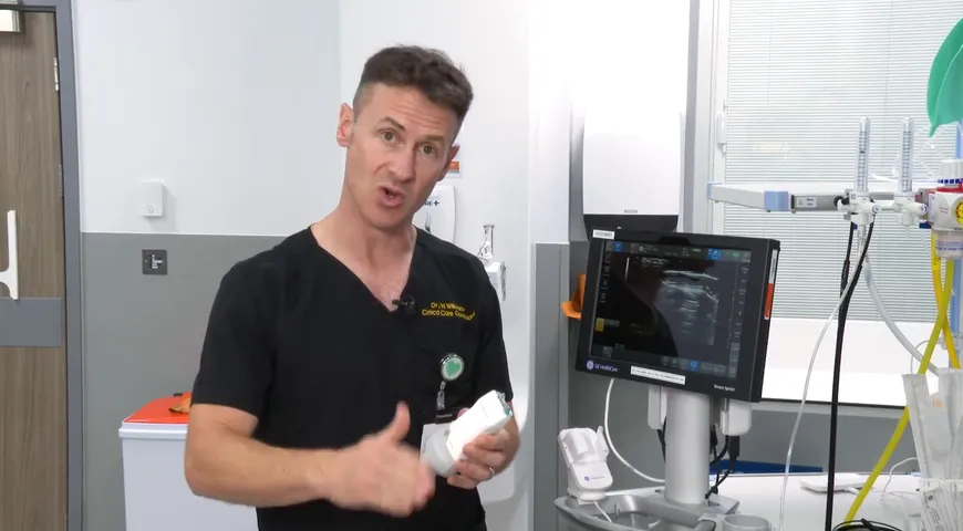
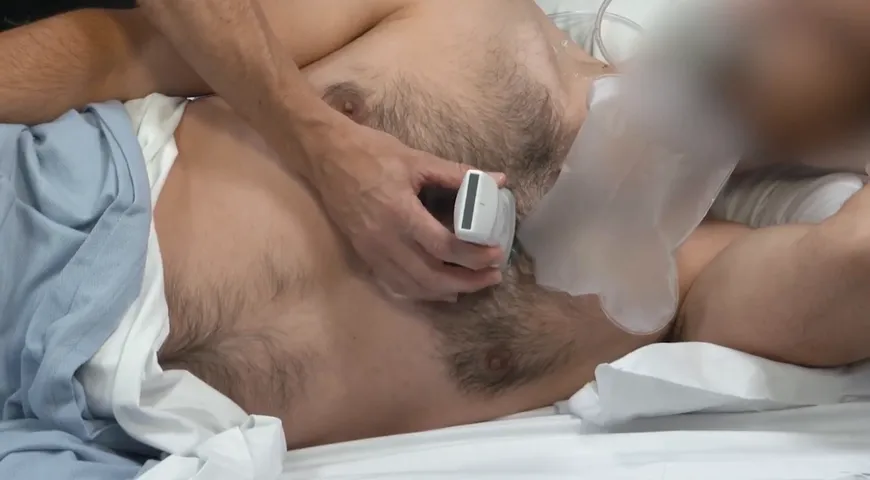
Dr Ashley Miller, a consultant intensivist from Shrewsbury and Telfords Hospital NHS Trust in the UK, teaches us how to perform an assessment of the lungs using ultrasound for an acutely unwell patient. A chest ultrasound is an assessment that produces images which can be used to provide a quick visualisation of the lungs. Dr. Miller takes us through which probe to use to get the best pictures and results, he suggests the curvilinear abdominal probe is the best for this type of assessment. Using three points of each side of the chest, the upper anterior, the lower anterior and the posteroanterior view. During the examination, you may have to ask you patient to hold their breath, to allow you see when functionality is stopped and what effect this has on the view you can see, such as thee lung pulse and lung sliding.  To learn more about this type of assessment watch the video in full for more information. 

Related

Lung Ultrasound Pearls
01:28 min

Lung Ultrasound Pearls

Dr. Ashley Miller

Contrast-Enhanced Ultrasound- Guided Biopsies at Odense University Hospital
04:03 min

Contrast-Enhanced Ultrasound- Guided Biopsies at Odense University Hospital

Prof. Christian B. Laursen

L’évaluation des fonctions VD et VG : aller plus loin que l’évaluation visuelle.
41:39 min

L’évaluation des fonctions VD et VG : aller plus loin que l’évaluation visuelle.

Prof. Laurent Muller

La ecografía en el tratamiento y seguimiento de paciente con dolor
04:24 min

La ecografía en el tratamiento y seguimiento de paciente con dolor

Dr. Alejandro Ortega

Echographie clinique du patient aigu & thrombose :  De la suspicion de thrombose veineuse prodonde à l'embolie pulmonaire grave
55:36 min

Echographie clinique du patient aigu & thrombose : De la suspicion de thrombose veineuse prodonde à...

Prof. Laurent Muller

De la décompensation cardiaque au choc cardiogégique
01:05:11 h

De la décompensation cardiaque au choc cardiogégique

Prof. Xavier Bobbia