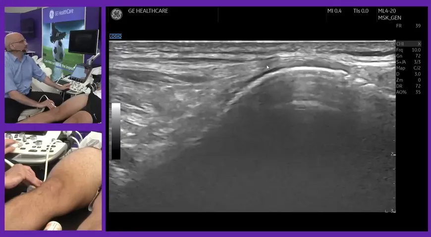
The purpose of this video is to identify appropriate probe positions when scanning the extensive compartments of the wrist. The demo is performed by Research Physiotherapist Mark Maybury who specialises in sonography.
Ultrasound probe positioning can be challenging and presents great difficulties in achieving good diagnostic quality ultrasounds. To ensure high-quality and easy-to-evaluate images, it essential that the probe is positioned correctly. Not being able to identify where you are scanning to seamlessly flow through the patient's wrist to each extensor compartment will result in a lengthy and low-resolution ultrasound.
The first extensor compartment is made up of the abductor pollicis longus and the extensor pollicis brevis. This area can be viewed by positioning the patient's hand on its side (semi-probnative/ sub-super probnative) and placing the probe above the wrist.
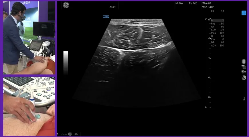
To view the second extensor compartment your patient's hand must be lying flat (pronated). The probe is then positioned on the patient's wrist. In this view, you should be able to see the extensor carpi radialis brevis and the extensor carpi radialis longus.
The third, fourth and fifth extensor compartment is viewed by placing the probe flat on the back of the patient's wrist.
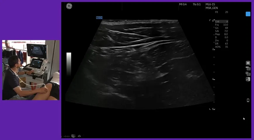
Finally, the sixth extensor compartment is examined by moving the probe laterally on the patient's wrist to view the extensor carpi ulnaris. While in this position, Mark moves the probe proximally to view the distal radioulnar joint.
Watch the full demonstration here.