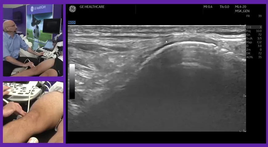
Mark Maybury is a Research Physiotherapist and Sonographer. In this video, he carries out a live demonstration of an ultrasound scanning of the flexor aspect of the wrist.
This video provides guidance on how to position your ultrasound probe on the patient's wrist to examine the flexor tendons, the median nerve, the ulnar nerve and the tendons that lead into the palm of the hand.
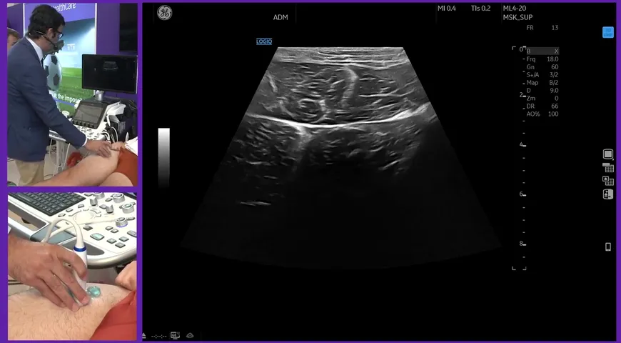
Ultrasound is an extremely valuable imaging tool to evaluate the wrist. It allows for high-quality imaging of the anatomy of the wrist, it also enables medical professionals to evaluate the tendons, ligaments and the wrist joint.
Being able to execute this procedure for your patients will allow you to easily diagnose such injuries as carpal tunnel syndrome, bursitis and joint effusion.
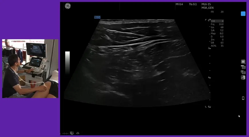
Watch Mark carry out a live demonstration of a wrist ultrasound and identify a number of nerves and tendons located around the first and lower forearm. For a step-by-step ultrasound demonstration, watch this flexor aspect video now.