Mark Maybury is a Research Physiotherapist and Sonographer. In this video, he carries out a live demonstration of an ultrasound of the long head of biceps tendon in the shoulder.
Watch the video to learn how to ultra-scan the long head of biceps tendon in the shoulder, position your probe and identifying different tendons surrounding the long head of biceps.
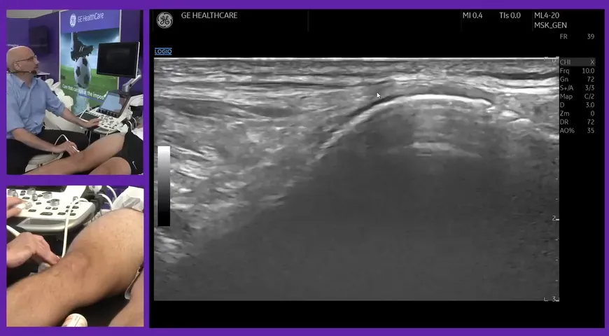
The long head of biceps tendon is looked at in a transverse view, positioning a probe vertically against the patient's bicipital groove. On the ultrasound, Mark points out the bicipital groove on the screen and points out other vital tendons in the shoulder.
Mark continues to evaluate the bicep tendon in different positions. It's important to evaluate the bicep tendon in many positions to make sure that the bicep is moving correctly and isn't jolting out or there aren't any tears.
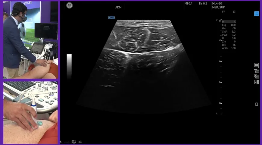
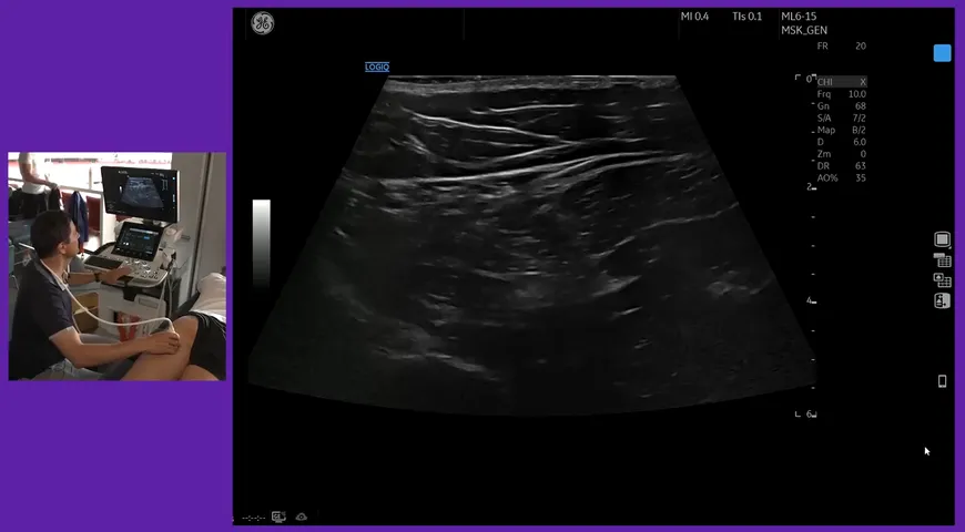
Mark then turns the probe 90 degrees laterally to view the bicep tendon in a long section view. The bicep tendon is now in a longitudinal view and allows you to further evaluate the tendon.
There are many studies to prove that shoulder ultrasound is an extremely accurate method to confirm a normal long head of biceps tendon or an injury such as a full-tear. For more information on this subject, please view the following video of how to ultrasound the shoulder.