Mark Maybury is a Research Physiotherapist Sonographer who will be taking part in a live demonstration video to explain how to perform an ultrasound of a shoulder.
Within this video, Mark goes into detail about the different tendons and joints that are located around the shoulder, and how you, a medical professional can identify them by positioning your probe in different areas around the shoulder.
Being able to perform an ultrasound of the shoulder is extremely beneficial because it can show any swelling around the joint and as well as any damage or problems with the tendons, muscles or any other kind of tissue around the shoulder.
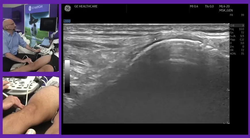
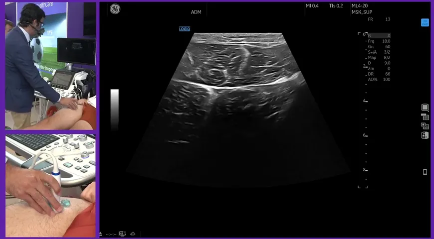
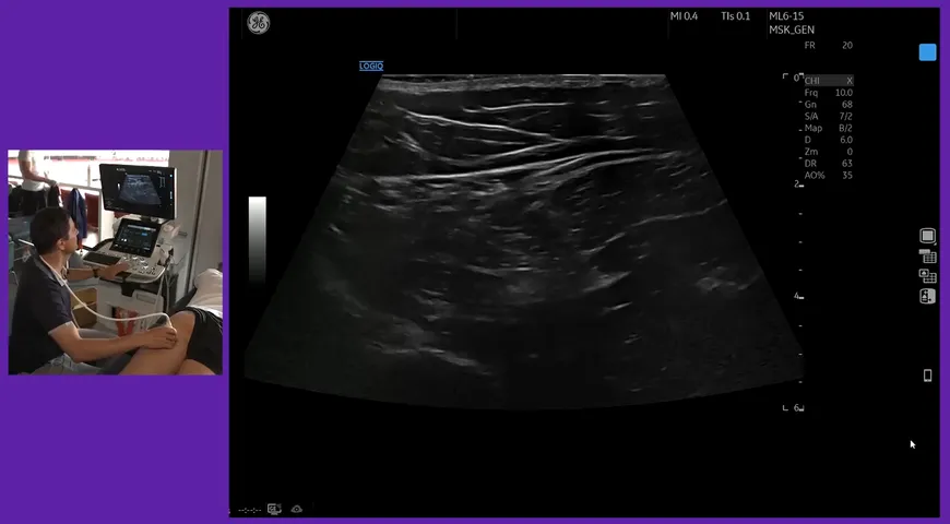
The most common muscles that are viewed in a shoulder ultrasound are the Subscapularis, Supraspinatus (between these two cuff muscles is where you will find the long head of bicep tendon), Infraspinatus and the Teres Minor. These four muscles make up the rotator cuff and each aid the motion of your shoulder.
The rotator cuff is one of the most common shoulder injuries that medical professions have to investigate, and it can be quite challenging to perform a shoulder ultrasound due to the makeup of the shoulder. However, in this video, Mark takes you through step-by-step how to perform a shoulder ultrasound, demonstrating all the different tendons and tissues. Watch the video below for more information on how to ultrasound a shoulder.