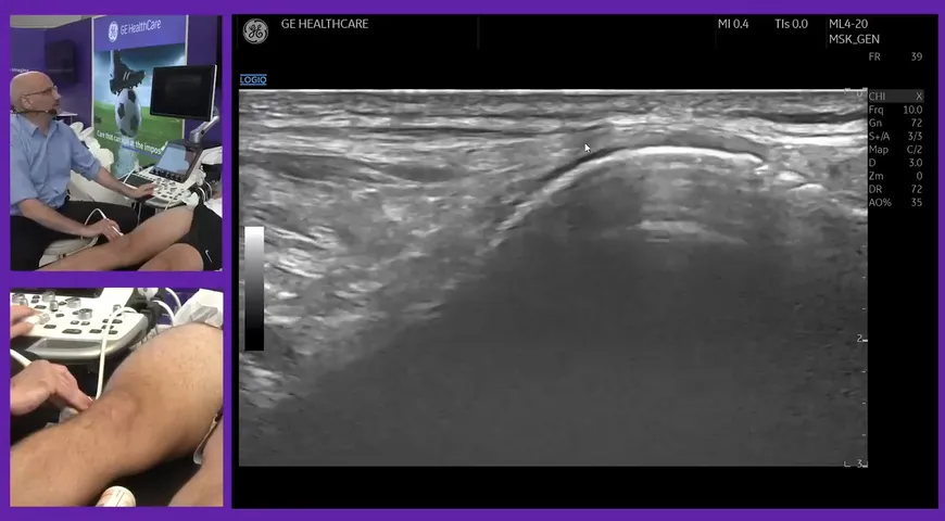
Mark Maybury, a Research Physiotherapist specialising in ultrasonography, demonstrates a live ultrasound of a distal bicep tendon; also known as the pronator teres view.
In this video clip, Mark utilises the pronator teres muscle as an acoustic window to look at the distal bicep tendon. He positions the patient's arm in a 90-degree flexion and places the ultrasound probe a proximity 5 cm above the olecranon is roughly where the distal bicep tendon sits.
This scan can be difficult to obtain but once mastered, this technique becomes an extremely useful view for medical professionals who are looking at the dynamic interplay between the distal bicep tendon. It provides information about whether or not the tendon is tendinopathic or whether there is an associated distal bicep bursa.
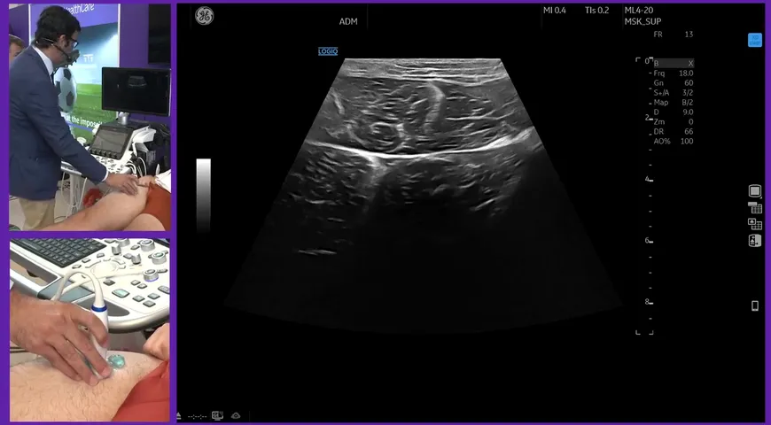
After Mark demonstrates how to position the probe on the patient, he goes on to discuss the ultrasound image that determines the brachial artery. This is a valuable anatomy structure to be able to identify on your screen because directly beneath it the distal bicep tendon.
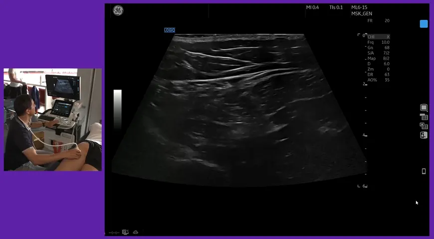
Mark then goes on to discuss the radial head and the cobra view. To watch the entire ultrasound demo of distal bicep tendon, click the link below.