Mark Maybury talks us through how to perform an ultrasound scan for the anterior elbow.
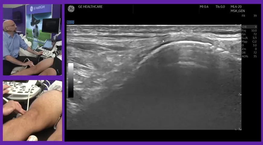
To begin, Mark places his probe high up on the anterior aspect of the patient's arm around the mid-shaft of the humerus.
At the top of the ultrasound screen, Mark points out the biceps which will form the central tendon of the distal bicep tendon. Underneath is brachialis and the humorous.
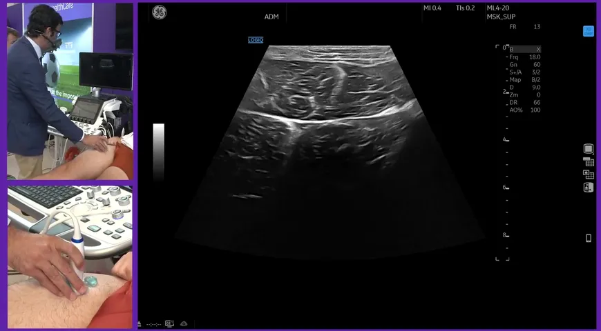
As Mark slides the probe down the patient's arm, the muscle section becomes smaller, and the tendon part becomes bigger. When he reaches the end, the muscle has coalesced to the bicep tendon. A little bit lower down there should be a clamp-like shape visible on the screen. This is the anterior joint line of the elbow: the articular cartilage and the cortical bone is present.
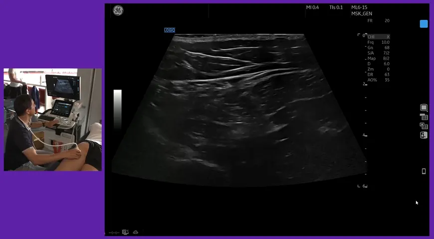
Mark continues to scan down the arm until the brachial artery is present. The bicep tendon goes dark and begins to turn onto its side. Mark then follows it down by switching to short side axis to its insertion on to radial tuberosity where it's no longer visible.
Watch the video below to learn more about the anterior aspect view of the elbow.