The following video will demonstrate how a medical professional should carry out an ultrasound of the cartilage of the ear to diagnosis polychondritis.
Professor Valentin Schafer MD is the head of the Department of Rheumatology & Clinical Immunology at the University Hospital in Bonn and is an expert in ultrasound imaging.
As a Rheumatologist, it's not uncommon to treat patients who are suffering from polychondritis which not only affects the ear but also other parts of the body.
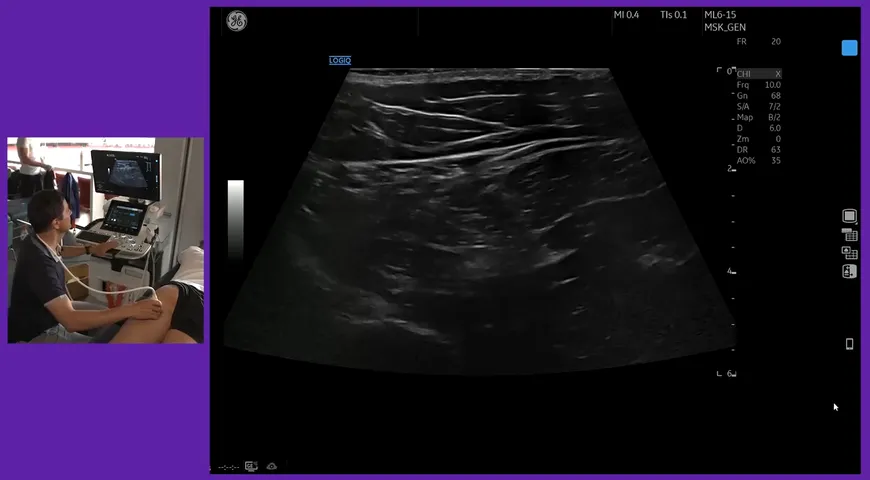
The ear is an easily diagnosed body part through ultrasound and in this video Professor Valentin will carry out a live demonstration of how to do this.
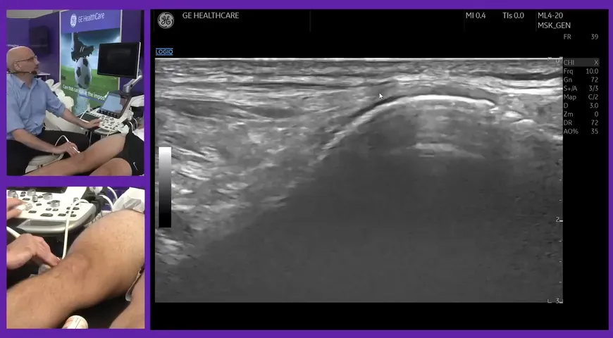
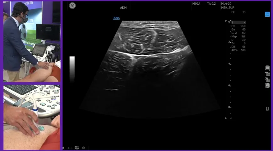
To start, Professor Valentin states that a high-resolution probe is needed. Before placing the probe on the patient's ear on top of the cartilag Professor Valentin continues to label the different anatomies of the image present and most importantly the cartilage.
Using ultrasound for the diagnosis of polychondritis is a quick and easy identifier without to much stress to the patient. To learn more about the diagnosis of polychondritis via ultrasound, watch this video.