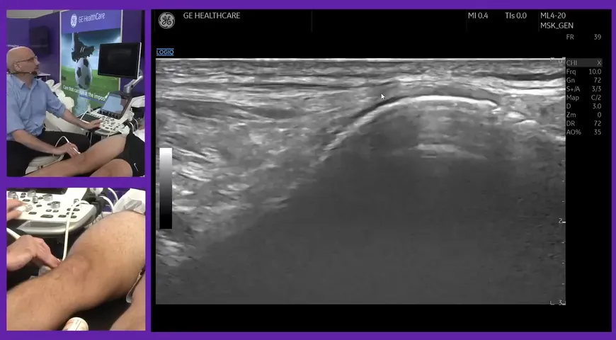
The axillary artery and ultrasound work hand in hand for the detection and diagnosis of Gaint-cell arthritis. In the following video, Professor Valentin Schafer MD who is the head of the Department of Rheumatology & Clinical Immunology at the University Hospital in Bonn will undertake a live performance of an ultrasound scan for the diagnosis of Gaint-cell arteritis (GCA).
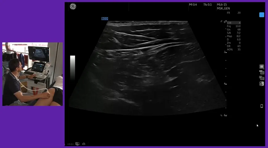
The beginning of the process involves a linear ultrasound probe (6-15 MHz) and placing it to the ear. This means you can see the temporal artery exit the skull. Professor Valentin then leads us through the artery as it splits into two arteries.
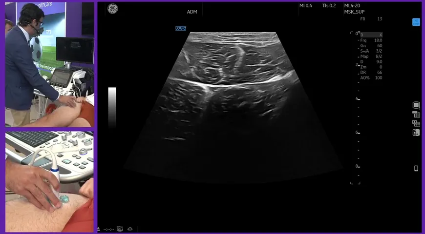
In this ultrasound scan, the patient has an anomaly in the common temporal artery. A useful tip that Professor Valentin shares with you is that if you have a lower Mhz ultrasound probe, you can use the compression technique to see whether or not the temporary artery can completely compress.
Watch the video above to learn more about GCA, the temporary artery, how to diagnosis GCA and to visually see how this patient's arteries compression, and what you should look for in order to give a correct diagnosis.
21