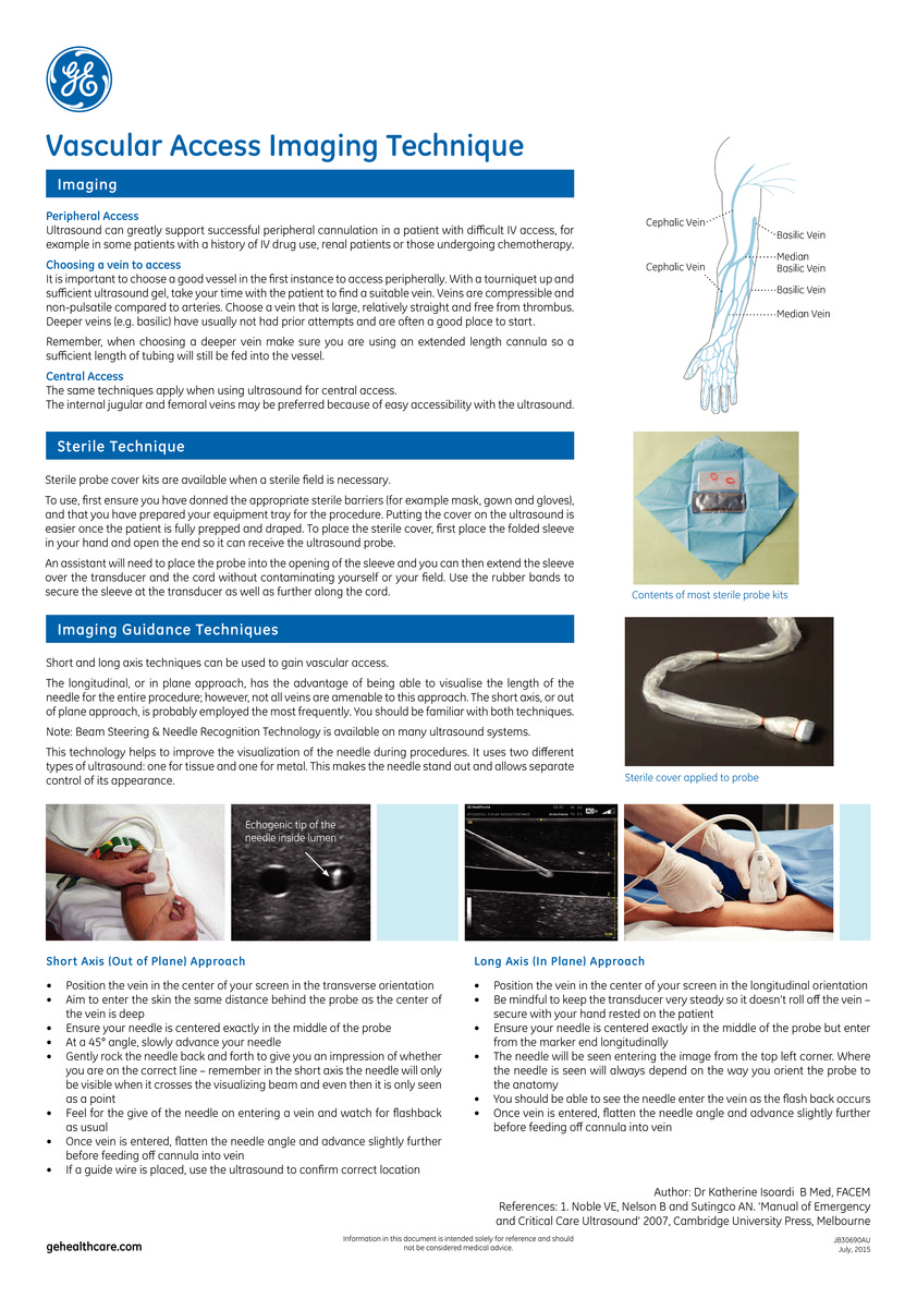
Vascular access through ultrasound can greatly support successful peripheral cannulation in a patient. It either reduces the number of PIV attempts or prevents the need for CVC insertion. Therefore, it is important that all providers be comfortable with this application, and aware of the best practices and techniques for improved performance with this procedure. This technique is especially important if you are dealing with a patient with difficult IV access, for example, those with a history of IV drug use or those undergoing chemotherapy. Being able to carry out IV ultrasound approach will mean that you can accurately determine the vein through ultra scanning, view exactly where you are piercing the sink, and successfully draw blood from the patient.
The use of ultrasound-guided vascular access improves placement success with a significant number of patients benefiting from this approach. Find out the benefits of vascular access imaging techniques below, the difference between imaging and sterile techniques and also how to carry out a short and long-axis imaging approach.
9