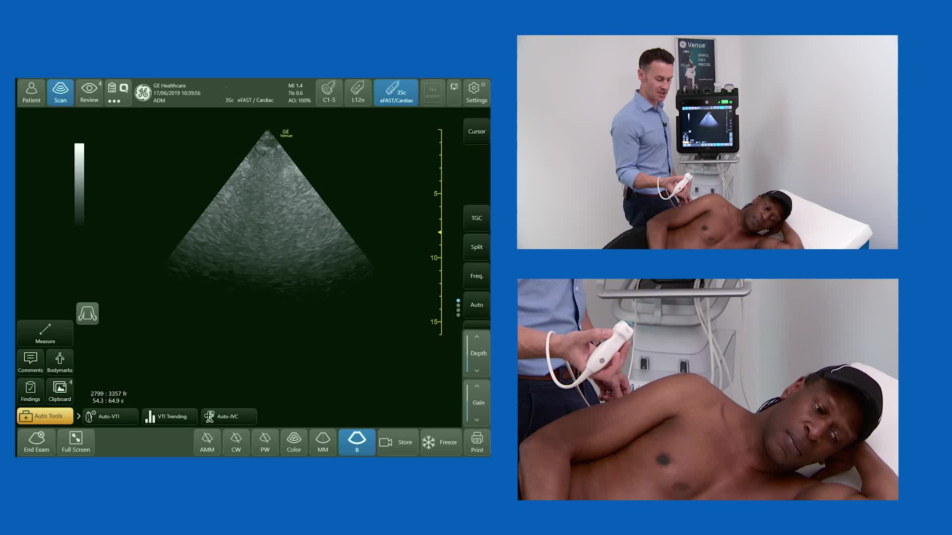
Dr. Jonny Wilkinson, Consultant in Intensive Care and Anaesthesia in Northampton, details in this short video on how to perform a basic cardiac ultrasound through looking at the parasternal long axis, the subcostal IVC, short axis and apical 4 and 5 chambers of the heart. A Cardiac ultrasound, also known as echocardiography, concerns the ultrasound imaging of a very fast moving complex organ positioned deep within the body. Cardiac ultrasound shows the size, structure, and movement of various parts of your patients heart. These parts include the heart valves, the septum (the wall separating the right and left heart chambers), and the walls of the heart chambers. He introduces this topic by performing this on a patient and asking them to lie on their left lateral to give you the optimal view set, he then takes us through the stages for performing this ultrasound. This is a step by step video that will give you the most optimal way to perform a basic cardiac ultrasound for you and your patient. To learn more about this type of assessment watch the video in full for more information.
27